Theanine and Caffeine: the yin-yang of green tea and brain waves

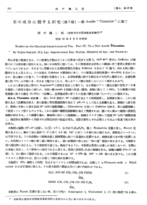
Theanine was discovered in 1949 by the Japanese researcher Yajiro Sakato1, as a new amide in the water extract of the Japanese green tea Gyokuro (玉 露) – also called “precious dew” due to its dark green colour and high price in Japanese markets, because of its unique characteristic taste of sweetness and “umami”2.

The green tea leaves from specialized varieties of the tea plant Camellia sinensis (Ericales plants) have enormous amounts of theanine, which they absorb from the roots of the plant depending on the nitrogen supply of the soil3, 4. A soil that is rich in nitrogen will promote the biosynthesis of the non-protein aminoacid L-Theanine from Glutamic Acid and Ethylamine (by the enzyme theanine synthetase)2. Since the commercial price of green tea is almost directly proportional to its theanine content, to obtain theanine-rich, good quality green tea leaves, a large amount of nitrogen fertilizer must be supplied to the cultivated plants throughout the growth period (with problematic effects on the environment)2.

Theanine is then transported via the xylem fluid, from the roots to the young bushes leaves. And, because light is necessary to the conversion of theanine into cathechins in the leaves of the plant, when Camellia sinensis bushes are protected from direct sunlight for a couple of weeks just before harvest, they have a high theanineamino acid content2. Even though cathechins are polyphenols with known antioxidant properties, they are also responsible for the astringent flavour of green tea. So, for an optimum taste, cathechins must be balanced with theanines. With less sunlight, there is less photosynthesis and leaf senescence; and, less theanine being converted into catechin, keeping the unique sweet-umami flavor characteristic of Gyokuro green tea.

Theanine is also known as N5-ethyl-L-glutamine due to its structural similarities to L-glutamic acid, which is the most abundant excitatory neurotransmitter in our brains5. Researchers think that L-theanine mechanism of action might be mediated by glutamate receptors, and it might act as a partial agonist for the N-methyl-D-aspartate receptor6

Theanine has known relaxing and anxiolytic effects, via the induction of the slow alpha-brain waves in the occipital and parietal regions of the human brain7. Plus, it doesn’t have any additive or side effects that are usually associated with conventional sleep inducers. 

There is only one IF…. 

In addition to L-TheanineCamellia sinensis leaves grown in the shade also have a high level of caffeine8, which decreases slow brain activity and keeps us awake (by increasing beta-wave activity). This because, the buds and young leaves of Camellia plants contain more caffeine than mature leaves8. As such, besides a high level of theanineGyokuro or Matcha green tea powder (which goes through the same shade process before harvest), also have high levels of caffeine

What is interesting, is that this dual effect of L-Theanine and Caffeine in Gyokuro or Matcha green tea powder, seem to have a synergistic effect in decreasing mind wandering and enhancing our attention to target stimuli9, 10. This was shown in a very small randomized clinical trial, that used functional Magnetic Resonance Imaging (fMRI) to scan the brains of subjects, after they ingested L-Theanine and Caffeine supplements while performing a visual task. 

So, if we want to focus and stay awake: a cup of Gyokuro or Matcha, will keep our attention sharp as a Japanese sword. 

If, on the other hand, we are not feeling very calm, anxiety is setting in, or if sleep is taking too long because the news are only “so-so” at the moment: 200-400mg of L-theanine could help us keep the zen mood, and have a good night sleep11.
The discovery of Theanine

References:

1.         Sakato Y. Studies on the Chemical Constituents of Tea

Part III. On a New Amide <b>Theanine</b>. Nippon Nōgeikagaku Kaishi. 1950;23:262-267.

2.         Ashihara H. Occurrence, biosynthesis and metabolism of theanine (γ-glutamyl-L-ethylamide) in plants: a comprehensive review. Nat Prod Commun. 2015;10:803-10.

3.         Ruan J, Haerdter R and Gerendás J. Impact of nitrogen supply on carbon/nitrogen allocation: a case study on amino acids and catechins in green tea [Camellia sinensis (L.) O. Kuntze] plants. Plant Biol (Stuttg). 2010;12:724-34.

4.         Huang H, Yao Q, Xia E and Gao L. Metabolomics and Transcriptomics Analyses Reveal Nitrogen Influences on the Accumulation of Flavonoids and Amino Acids in Young Shoots of Tea Plant ( Camellia sinensis L.) Associated with Tea Flavor. J Agric Food Chem. 2018;66:9828-9838.

5.         Unno K, Furushima D, Hamamoto S, Iguchi K, Yamada H, Morita A, Horie H and Nakamura Y. Stress-Reducing Function of Matcha Green Tea in Animal Experiments and Clinical Trials. Nutrients. 2018;10:1468.

6.         Sebih F, Rousset M, Bellahouel S, Rolland M, de Jesus Ferreira MC, Guiramand J, Cohen-Solal C, Barbanel G, Cens T, Abouazza M, Tassou A, Gratuze M, Meusnier C, Charnet P, Vignes M and Rolland V. Characterization of l-Theanine Excitatory Actions on Hippocampal Neurons: Toward the Generation of Novel N-Methyl-d-aspartate Receptor Modulators Based on Its Backbone. ACS Chem Neurosci. 2017;8:1724-1734.

7.         Kobayashi K, Nagato Y, Aoi N, Juneja LR, Kim M, Yamamoto T and Sugimoto S. Effects of L-Theanine on the Release of &alpha;-Brain Waves in Human Volunteers. Nippon Nōgeikagaku Kaishi. 1998;72:153-157.

8.         Ashihara H and Suzuki T. Distribution and biosynthesis of caffeine in plants. Front Biosci. 2004;9:1864-76.

9.         Kahathuduwa CN, Dhanasekara CS, Chin SH, Davis T, Weerasinghe VS, Dassanayake TL and Binks M. l-Theanine and caffeine improve target-specific attention to visual stimuli by decreasing mind wandering: a human functional magnetic resonance imaging study. Nutr Res. 2018;49:67-78.

10.       Hidese S, Ogawa S, Ota M, Ishida I, Yasukawa Z, Ozeki M and Kunugi H. Effects of L-Theanine Administration on Stress-Related Symptoms and Cognitive Functions in Healthy Adults: A Randomized Controlled Trial. Nutrients. 2019;11:2362.

11.       Williams JL, Everett JM, D’Cunha NM, Sergi D, Georgousopoulou EN, Keegan RJ, McKune AJ, Mellor DD, Anstice N and Naumovski N. The Effects of Green Tea Amino Acid L-Theanine Consumption on the Ability to Manage Stress and Anxiety Levels: a Systematic Review. Plant Foods Hum Nutr. 2020;75:12-23.

Feeling anxious or depressed? Might be your microglia…

A macrophage is a hungry immune cell that engulfs and eats all things that don’t have a good reputation in our body (e.g., cellular debris, pathogens…); and, microglia cells are the resident macrophage population of the Central Nervous System (CNS)1. They function as sentinels of local infection in the brain, backing both innate and adaptive immune responses, and account for 10-15% of all cells found in the brain and spinal cord2.

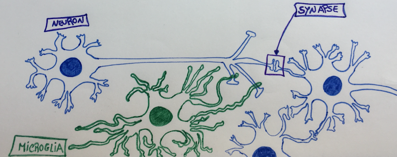
Microglia cells are also involved in the maintenance of brain homeostasis, contributing to mechanisms that underly learning and memory. They constantly survey their local microenvironment – like patrols – extending their motile processes, or hands/legs, to make a brief contact with neuronal synapses. This continuous synaptic plasticity, throughout our lifetime, is essential to control maladaptive learning and memory, such as addiction3. For example, the number of synapses in the brain regions of the nucleus accumbensamygdala and dorsomedial striatum increase when we expose our brains to addictive substances (such as alcohol, or opiates); and, decrease upon withdrawal due to the action of microglia cells4. As such, microglia cells help to modify and eliminate synaptic structures when they grow too much, or, are on the way to touch too many other neurons5 – because, neurons tend to be touchy and to enjoy a synaptic orgy. 

Whenever a neuron starts to freak out that it has too many synapses and it needs help regulating its neuronal “touchy” behaviour, then the synapse extends a greeting “hand” (filopodia) and “Hi5s” the neighbouring microglia cell, telling her that it needs help remodelling. Once “Hi5ed”, the microglia cell starts nibbling on the synapse6 – cutting all the excess – and, avoiding that that specific neuron gets assigned a bad “sexual” reputation. It’s like behaviour counselling, transforming and remodelling, but neuron-wise and with a microglia cell as the counsellor…

Even though microglia cells are essential and extremely helpful; like everything in life, they can also go haywire, ending up pruning too many synapses, and destroying healthy tissue. An uncontrolled activation of the microglia can be directly toxic to neurons, because they can release inflammatory cytokines (IL-1, TNF-alpha, IL-6, Nitric Oxide, Prostaglandine E2, and Superoxide)7, and lead to excessive pruning of neuronal synapses3.

The most recent research in the pathophysiology of depression and anxiety shows that abnormalities in microglia cells have a central role in the development of these diseases8. For example, a neuroimaging study in depressed patients, revealed that stronger depressive symptoms related with microglial activation in brain regions associated with mood regulation (the prefrontalanterior cingulate, and insular cortices of the brain)9. Additionally, post-mortem studies of depressed suicide victims showed microglial activation and macrophage accumulation within the anterior cingulate cortex brain region10

Persistent stress activates a chronic low-inflammatory state in our bodies that enhances our inflammatory response to challenges11. Social stress causes the release of inflammatory monocytes into the circulation8, which end up reaching the Blood Brain Barrier (BBB) and its endothelial cells. This low-systemic inflammation that travels through our vessels, encourages the migration of the brain resident microglia cells to the area of the cerebral vessels. In here, microglia cells make physical contact with endothelial cells of the BBB, and “sense” the inflammatory environment that is present in the blood (aka, inflammatory cytokines activate receptors in the microglia cells). If there is sustained inflammation, then some of the microglia cells can “become neurotic” and start nibbling the end-feet of healthy cells, making the BBB more permeable and, consequently, damaging the protective BBB shield function12. This is turn, leaks inflammatory cytokines from the blood into the brain tissue, further activating more microglia cells, that start cutting synapses from healthy neurons.

What this means is that a persistent low-grade inflammation can trigger microglia activation and change the functional connectivity of healthy neurons in major brain emotional centers13. Because our immune system can interact with the neurocircuitry that is involved in emotion regulation and behaviour, a chronic low-inflammation derived from stress can influence the development of various neuropsychiatric disorders, like depression and anxiety. 

But, what can we do to avoid falling in this trap?

Eat well, sleep well, do sports and have a good laugh with friends. All things that inhibit inflammation, and make us feel good. 

Microglia cell (green) “counselling” a synapse

References:

1.         Ginhoux F, Lim S, Hoeffel G, Low D, Huber T. Origin and differentiation of microglia. Frontiers in Cellular Neuroscience. 2013;7

2.         Lawson LJ, Perry VH, Gordon S. Turnover of resident microglia in the normal adult mouse brain. Neuroscience. 1992;48:405-415

3.         Neniskyte U, Gross CT. Errant gardeners: Glial-cell-dependent synaptic pruning and neurodevelopmental disorders. Nat Rev Neurosci. 2017;18:658-670

4.         Spiga S, Talani G, Mulas G, Licheri V, Fois GR, Muggironi G, et al. Hampered long-term depression and thin spine loss in the nucleus accumbens of ethanol-dependent rats. Proc Natl Acad Sci U S A. 2014;111:E3745-3754

5.         Tremblay M-È, Lowery RL, Majewska AK. Microglial interactions with synapses are modulated by visual experience. PLOS Biology. 2010;8:e1000527

6.         Weinhard L, di Bartolomei G, Bolasco G, Machado P, Schieber NL, Neniskyte U, et al. Microglia remodel synapses by presynaptic trogocytosis and spine head filopodia induction. Nature Communications. 2018;9:1228

7.         Kim YS, Joh TH. Microglia, major player in the brain inflammation: Their roles in the pathogenesis of parkinson’s disease. Exp Mol Med. 2006;38:333-347

8.         McKim DB, Weber MD, Niraula A, Sawicki CM, Liu X, Jarrett BL, et al. Microglial recruitment of il-1β-producing monocytes to brain endothelium causes stress-induced anxiety. Mol Psychiatry. 2018;23:1421-1431

9.         Setiawan E, Wilson AA, Mizrahi R, Rusjan PM, Miler L, Rajkowska G, et al. Role of translocator protein density, a marker of neuroinflammation, in the brain during major depressive episodes. JAMA Psychiatry. 2015;72:268-275

10.       Suzuki H, Ohgidani M, Kuwano N, Chrétien F, Lorin de la Grandmaison G, Onaya M, et al. Suicide and microglia: Recent findings and future perspectives based on human studies. Frontiers in cellular neuroscience. 2019;13:31-31

11.       Miller GE, Rohleder N, Cole SW. Chronic interpersonal stress predicts activation of pro- and anti-inflammatory signaling pathways 6 months later. Psychosom Med. 2009;71:57-62

12.       Haruwaka K, Ikegami A, Tachibana Y, Ohno N, Konishi H, Hashimoto A, et al. Dual microglia effects on blood brain barrier permeability induced by systemic inflammation. Nature communications. 2019;10:5816-5816

13.       Kim J, Yoon S, Lee S, Hong H, Ha E, Joo Y, et al. A double-hit of stress and low-grade inflammation on functional brain network mediates posttraumatic stress symptoms. Nature Communications. 2020;11:1898