
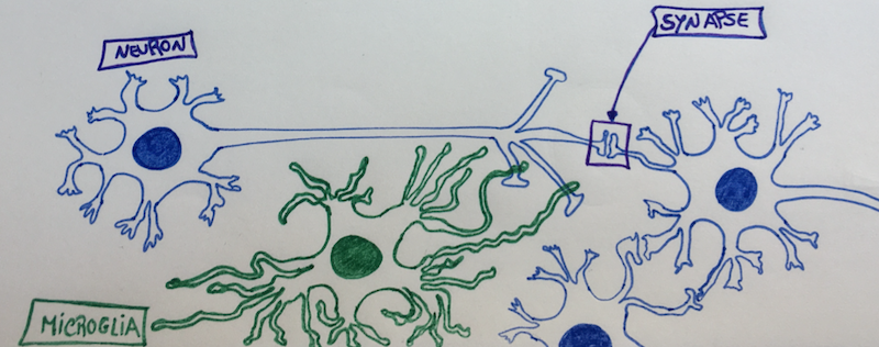
A macrophage is a hungry immune cell that engulfs and eats all things that don’t have a good reputation in our body (e.g., cellular debris, pathogens…); and, microglia cells are the resident macrophage population of the Central Nervous System (CNS)1. They function as sentinels of local infection in the brain, backing both innate and adaptive immune responses, and account for 10-15% of all cells found in the brain and spinal cord2.
Microglia cells are also involved in the maintenance of brain homeostasis, contributing to mechanisms that underly learning and memory. They constantly survey their local microenvironment – like patrols – extending their motile processes, or hands/legs, to make a brief contact with neuronal synapses. This continuous synaptic plasticity, throughout our lifetime, is essential to control maladaptive learning and memory, such as addiction3. For example, the number of synapses in the brain regions of the nucleus accumbens, amygdala and dorsomedial striatum increase when we expose our brains to addictive substances (such as alcohol, or opiates); and, decrease upon withdrawal due to the action of microglia cells4. As such, microglia cells help to modify and eliminate synaptic structures when they grow too much, or, are on the way to touch too many other neurons5 – because, neurons tend to be touchy and to enjoy a synaptic orgy.
Whenever a neuron starts to freak out that it has too many synapses and it needs help regulating its neuronal “touchy” behaviour, then the synapse extends a greeting “hand” (filopodia) and “Hi5s” the neighbouring microglia cell, telling her that it needs help remodelling. Once “Hi5ed”, the microglia cell starts nibbling on the synapse6 – cutting all the excess – and, avoiding that that specific neuron gets assigned a bad “sexual” reputation. It’s like behaviour counselling, transforming and remodelling, but neuron-wise and with a microglia cell as the counsellor…
Even though microglia cells are essential and extremely helpful; like everything in life, they can also go haywire, ending up pruning too many synapses, and destroying healthy tissue. An uncontrolled activation of the microglia can be directly toxic to neurons, because they can release inflammatory cytokines (IL-1, TNF-alpha, IL-6, Nitric Oxide, Prostaglandine E2, and Superoxide)7, and lead to excessive pruning of neuronal synapses3.
The most recent research in the pathophysiology of depression and anxiety shows that abnormalities in microglia cells have a central role in the development of these diseases8. For example, a neuroimaging study in depressed patients, revealed that stronger depressive symptoms related with microglial activation in brain regions associated with mood regulation (the prefrontal, anterior cingulate, and insular cortices of the brain)9. Additionally, post-mortem studies of depressed suicide victims showed microglial activation and macrophage accumulation within the anterior cingulate cortex brain region10.
Persistent stress activates a chronic low-inflammatory state in our bodies that enhances our inflammatory response to challenges11. Social stress causes the release of inflammatory monocytes into the circulation8, which end up reaching the Blood Brain Barrier (BBB) and its endothelial cells. This low-systemic inflammation that travels through our vessels, encourages the migration of the brain resident microglia cells to the area of the cerebral vessels. In here, microglia cells make physical contact with endothelial cells of the BBB, and “sense” the inflammatory environment that is present in the blood (aka, inflammatory cytokines activate receptors in the microglia cells). If there is sustained inflammation, then some of the microglia cells can “become neurotic” and start nibbling the end-feet of healthy cells, making the BBB more permeable and, consequently, damaging the protective BBB shield function12. This is turn, leaks inflammatory cytokines from the blood into the brain tissue, further activating more microglia cells, that start cutting synapses from healthy neurons.
What this means is that a persistent low-grade inflammation can trigger microglia activation and change the functional connectivity of healthy neurons in major brain emotional centers13. Because our immune system can interact with the neurocircuitry that is involved in emotion regulation and behaviour, a chronic low-inflammation derived from stress can influence the development of various neuropsychiatric disorders, like depression and anxiety.
But, what can we do to avoid falling in this trap?
Eat well, sleep well, do sports and have a good laugh with friends. All things that inhibit inflammation, and make us feel good.
References:
1. Ginhoux F, Lim S, Hoeffel G, Low D, Huber T. Origin and differentiation of microglia. Frontiers in Cellular Neuroscience. 2013;7
2. Lawson LJ, Perry VH, Gordon S. Turnover of resident microglia in the normal adult mouse brain. Neuroscience. 1992;48:405-415
3. Neniskyte U, Gross CT. Errant gardeners: Glial-cell-dependent synaptic pruning and neurodevelopmental disorders. Nat Rev Neurosci. 2017;18:658-670
4. Spiga S, Talani G, Mulas G, Licheri V, Fois GR, Muggironi G, et al. Hampered long-term depression and thin spine loss in the nucleus accumbens of ethanol-dependent rats. Proc Natl Acad Sci U S A. 2014;111:E3745-3754
5. Tremblay M-È, Lowery RL, Majewska AK. Microglial interactions with synapses are modulated by visual experience. PLOS Biology. 2010;8:e1000527
6. Weinhard L, di Bartolomei G, Bolasco G, Machado P, Schieber NL, Neniskyte U, et al. Microglia remodel synapses by presynaptic trogocytosis and spine head filopodia induction. Nature Communications. 2018;9:1228
7. Kim YS, Joh TH. Microglia, major player in the brain inflammation: Their roles in the pathogenesis of parkinson’s disease. Exp Mol Med. 2006;38:333-347
8. McKim DB, Weber MD, Niraula A, Sawicki CM, Liu X, Jarrett BL, et al. Microglial recruitment of il-1β-producing monocytes to brain endothelium causes stress-induced anxiety. Mol Psychiatry. 2018;23:1421-1431
9. Setiawan E, Wilson AA, Mizrahi R, Rusjan PM, Miler L, Rajkowska G, et al. Role of translocator protein density, a marker of neuroinflammation, in the brain during major depressive episodes. JAMA Psychiatry. 2015;72:268-275
10. Suzuki H, Ohgidani M, Kuwano N, Chrétien F, Lorin de la Grandmaison G, Onaya M, et al. Suicide and microglia: Recent findings and future perspectives based on human studies. Frontiers in cellular neuroscience. 2019;13:31-31
11. Miller GE, Rohleder N, Cole SW. Chronic interpersonal stress predicts activation of pro- and anti-inflammatory signaling pathways 6 months later. Psychosom Med. 2009;71:57-62
12. Haruwaka K, Ikegami A, Tachibana Y, Ohno N, Konishi H, Hashimoto A, et al. Dual microglia effects on blood brain barrier permeability induced by systemic inflammation. Nature communications. 2019;10:5816-5816
13. Kim J, Yoon S, Lee S, Hong H, Ha E, Joo Y, et al. A double-hit of stress and low-grade inflammation on functional brain network mediates posttraumatic stress symptoms. Nature Communications. 2020;11:1898