Psilocybin & Psilocin: Serotonin’s funny cousins

or the Magic Mushrooms Keychain

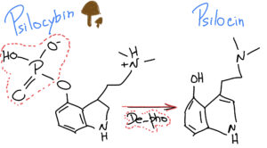
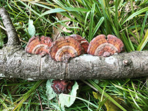
“Magic mushrooms” are fungi that contain Psylocibin

When we ingest Psylocibin, it gets degraded by the acid juices of our stomachs and loses its phosphate group (P), giving rise to a compound called Psilocin.

Psilocybin de-phosphorylation to Psilocin @countlesssheep.com
Psilocybin de-phosphorylation to Psilocin

What is interesting is that Psilocin (organic name: 4-hydroxy-N,N-dimetiltryptamine) is very similar to Serotonin – a very important neurotransmitter involved in our mood, learning and a plethora of other fundamental physiological processes.

Psilocin & Serotonin: similarities and differences @countlesssheeo.com
Psilocin & Serotonin: similarities and differences

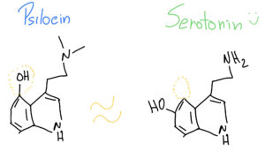
As such, when Psilocin reaches our prefrontal cortex, it can easily bind to our serotonin receptors, because they look so similar – it’s like two old keys that look almost alike and can open the same door at our grandmother’s house.

But Psilocin and Serotonin are indeed different. 

As such, Psilocin is able to do certain things in our brains when it binds to the similar Serotonin “door lock”, causing hallucinations and emotional changes that seem to alter the perception of space and time. Psilocin is like that funny cousin that can create chaos when it comes to visit during summer vacations… And, not all trips seem to be good trips, because as all things in life, a lot depends on the surrounding environment and the dosage. So, if a person comes through that door in a poor state of mind, it will just go down the “dark-hole” even further – so they say…

In fact a couple of years back, psychopharmacologists Robin Carhart-Harris and David Nutt from the Imperial College London did a fMRI study (functional magnetic resonance imaging) to evaluate the effects of Psilocybin in the brain2, and decided to give it IV (intravenously) to quicken the trip-effect because they were scared the “voyage” inside of the tight-noisy fMRI machine could be scary for the 30 individuals high on Psilocybin3. The results were quite interesting and showed that the effects of this psychedelic drug could be caused by a decreased activity and connectivity in the brain’s key connector hubs, like enabling a state of unconstrained cognition3. It seems  Psilocybin reduced the blood flow and neural activity in the posterior cingulate cortex and medial prefrontal cortex – almost like making a “software reset” of the brain. 

As such, Psilocin has been considered a serotonergic psychedelic compound; and it has been banned since the 70’s because people at the time thought it had no therapeutic value.

But Psilocin seems to have an effect in the treatment of Major Depressive Disorder (MDD), a leading cause of disability worldwide. Robin von Rotz and team at the Neurophenomenology of Conscious Lab from the University of Zürich, Switzerland, have just released the results of a randomized double-blind clinical trial 1. This clinical study showed that a single, moderate dose of Psilocybin (0.215 mg/Kg) significantly reduced depressive symptoms compared to a placebo, the “sugar-pill” that they give to the control group.

Even though the results were only evaluated for a period of two weeks after ingestion, the depression severity scores significantly improved in the treated patients in comparison with controls. So, this is one of the first clinical studies to actually demonstrate improvements directly attributed to Psilocybin/Psilocin itself. If we think that the state of deep depression is actually a neural circuitry disfunction, then Psilocin with its similar key structure to Serotonin might be able to open and clean the faulty neuronal-wires that contribute to the brain disfunction seen in MDD.

Several larger clinical studies are currently under way that could eventually pave the way to full regulatory approval, and the removal of Psilocybin from the banned WHO list of pure psychedelic drugs. The U.S. Food and Drug Administration already gave psilocybin the “breakthrough therapy” designation for MDD and Treatment-Resistant Disorder; and, in Australia some psychiatrist can have permission to use it under certain conditions for Post-Traumatic Stress Disorder. The European Medicines Agency (EMA) is following suit, and its Chief Medical Officer has just released a statement that it is actively engaged with developers of psychedelic therapies and academic researchers to help them identify what it takes to move forward and fully bring psychedelics as medical therapies to our pharmacies (and not street dealers)4.

We will be on the lookout for those results…

Fan shape mushrooms

References:

1          von Rotz, R. et al. Single-dose psilocybin-assisted therapy in major depressive disorder: a placebo-controlled, double-blind, randomised clinical trial. eClinicalMedicine 56 (2023). https://doi.org:10.1016/j.eclinm.2022.101809

2 Carhart-Harris, R. L. et al. The administration of psilocybin to healthy, hallucinogen-experienced volunteers in a mock-functional magnetic resonance imaging environment: a preliminary investigation of tolerability. J Psychopharmacol 25, 1562-1567 (2011). https://doi.org:10.1177/0269881110367445

3        Miller, G. Mapping the psychadelic brain. Science Brain & Behaviour (2012). https://doi.org:10.1126/article.27824

4   https://www.linkedin.com/pulse/second-chance-psychedelics-european-medicines-agency

Scleractinia & Symbiodinium: a tragic love story

The exchange of small molecules, or metabolites, between organisms and their environment gives rise to the complexity of life on Earth1. One of the examples of such complexity is ourselves, our body and the full array of microorganisms (microbiota) that live on and in us, humans. 

Just like us, stony corals (Scleractinia) also live in a symbiosis with associated microorganisms. These include bacteria, archea, fungi, viruses, and single-cellular algae called Symbiodinium.

Under normal conditions, these algal cells provide 90 to 95% of the energy required for stony corals to live happily1. The algae provide lipids, carbohydrates, amino acids, and O2 to Scleractinia. In return, the algae get to recycle nitrogen and other inorganic compounds from the coral, that they can use to fuel their own cell metabolism.

Climate change causes a rise in water temperatures; and, this form of environmental heat stress, disrupts the symbiotic relationship between corals and its algae, which results in coral bleaching. 

Researchers have now discovered that heat stress destabilizes this basic nutrient exchange between corals and their algae; and, this turn of events happens long before the bleaching process becomes obvious. 

In fact, Rädecker and his colleagues2 at the Red Sea Research Centre, discovered that heat stress causes the coral to need more energy, which swings the coral-algae symbiosis from a nitrogen- to a carbon-limited state. This actually ends up reducing the movement and recycling of carbon, and creates an animosity between these life partners. 

The coral starts disliking the algae, because the algae it’s not fully doing its work – recycling carbon. As such, the stony coral starts thinking that sheltering such lazy algae is not beneficial anymore. On top of that, the poor coral needs to catabolically degrade more aminoacids, to compensate for the increased energy demands triggered by the warmer waters. Such demanding “cooking tasks” from the coral side, create fury, and actually increase the release of ammonium; which in turn, promote the algae to grow. So, not only does the algae not clean the building properly, but calls its algal friends and they move in. 

Under stress, the coral kicks the algae out. But since this marriage is essential to its life support, the Scleractinia cannot feed itself properly anymore. The stony coral goes hungry, and slowly dies out….

Poor coral…

Underwater love

References:

1          Williams, A. et al. Metabolomic shifts associated with heat stress in coral holobionts. Science Advances 7, eabd4210, doi:doi:10.1126/sciadv.abd4210 (2021).

2          Rädecker, N. et al. Heat stress destabilizes symbiotic nutrient cycling in corals. Proceedings of the National Academy of Sciences 118, e2022653118, doi:10.1073/pnas.2022653118 (2021).

The language of serotonin

Or, “What are they saying?

When we mention the word Serotonin (5-hydroxytryptamine, 5-HT), we immediately think of the brain and the Central Nervous System (CNS). People tend to associate serotonin to depression, or mood, or feelings of well-being1

Although that is correct, truth be told, the majority of the serotonin in the human body is actually produced in the gut. In fact, 95% of total serotonin is manufactured by the Enterochromaffin cells (or, Kulchitsky cells) in the gastro-intestinal tract (GI)2,3. These cells live next to the gut epithelium, that covers the cavity of the GI tract, playing a crucial role in the regulation of bowel movements and secretions. If you think that the gut is almost 9 meters (or 30 feet) long, then that’s a lot of cells producing serotonin. 

When in the 50’s, Betty M. Twarog and Irvine H. Page discovered that the brain produced its own serotonin4; then, the gut-made serotonin got reduced to its “Aschenputtel” origins, and relinquished to the favela quarters of the body. As such, brain-derived serotonin always got more attention than its gut-derived counterpart – like a rich vs. poor-cousin type of reputation.

Moving-on…

Platelets, also called thrombocytes, are small un-nucleated fragment of cells that, when activated, form blood clots (thrombus) and prevent bleeding. 

Electron microscopy images of circulating platelets, extracted from Zilla et al, 19875

Platelets do not make serotonin, butcan take it up as they circulate through the gut, and carry it along the blood stream6,7. As such, the serotonin produced in the intestine can be carried all over the body. As the chemical messenger serotonin is, it can influence any other cell, in whatever other location, as long as it has a serotonin receptor on it. As such, peripheral serotonin has now discovered its path back into the limelight, and recent research has strengthened the influence that gut-made serotonin has in other parts of the body, functioning as an intestinal-derived hormone. 

Once again, the “Aschenputtel” story comes into mind, but this time through its “Cinderella” version. Let’s take a look…

For example, gut-derived serotonin can directly regulate the liver and mediate liver regeneration8. In Non-Alcoholic Fatty Liver Disease (NAFLD), a group of conditions that are characterized by excessive fat accumulation in the liver and closely track the global public health problem of obesity, researchers showed that inhibiting gut-derived serotonin synthesis could resolve hepatic fat accumulation8,9.

Peripheral serotonin can also be a negative regulator of bone density, by specifically inhibiting osteoblast formation and leading to osteoporosis10 – a common feature in patients with inflammatory bowel disease (IBD). This happens through the action of a common receptor: the low-density Lipoprotein Receptor-related Protein 5(LRP5), which is expressed in both osteoblasts and enterochromaffin cells11. LRP5 inhibits the expression of an important ingredient for serotonin production (Tryptophan hydroxylase-1, Tph1); as such, when LRP5 is deficient or inactivated due to inflammation or disease, blood levels of serotonin are elevated decreasing osteoblast formation; and, consequently, reducing bone mass1,11.

Epidemiologic data suggests a role of serotonin, or Selective Serotonin-Reuptake Inhibitors (typically used as antidepressants, SSRIs) in the development of venous thrombosis12. In fact, patients with depression were reported to have higher incidences of venous thromboembolism in general13; and, the use of SSRIs is associated with an increased venous thromboembolism risk14. No wonder, serotonin and platelets are “brothers in arms”, ready to block any blood vessel along their way…. 

Serotonin and its receptors are also present in the immune system, where evidence suggests it contributes to both innate and adaptive responses. There is now clear evidence of a straight communication between the immune system, the gut and the brain via serotonin15,16.

On top of all and because we are not alone, our gut microbiota plays a critical role in regulating our colonic serotonin. Indigenous spore-forming bacteria (Sp) promote serotonin biosynthesis in our enterochromaffin cells, and with that they can significantly modulate GI movements and platelet function – together with many aspects of our physiology17,18. We now know that the microbiota colonizes the GI tract after birth, with a continuous maturation during the first years of life19. Researchers have now showed in animal models that this developing gut microbiota regulates the development of the adult enteric nervous system via intestinal serotonin networks20. What this actually means, is that the actions of our intestinal bugs during the beginning of our life are determinant for the development of our “gut brain”, our second brain. How about that?…

If we ruminate about it, when we “think” with our gut, we are actually listening to our bugs. By directly signalling our cells to produce serotonin and develop a network of neurons as soon as we are born, our gut-bugs are actually finding a way to communicate with us – the host – in the serotonin language. 

Now, we just need to understand what are they telling us… 

Beethoven’s hearing aids, Beethoven House Museum, Bonn.

References:

1          Gershon, M. D. 5-Hydroxytryptamine (serotonin) in the gastrointestinal tract. Curr Opin Endocrinol Diabetes Obes 20, 14-21, doi:10.1097/MED.0b013e32835bc703 (2013).

2          Bellono, N. W. et al. Enterochromaffin Cells Are Gut Chemosensors that Couple to Sensory Neural Pathways. Cell 170, 185-198.e116, doi:10.1016/j.cell.2017.05.034 (2017).

3          Yaghoubfar, R. et al. Modulation of serotonin signaling/metabolism by Akkermansia muciniphila and its extracellular vesicles through the gut-brain axis in mice. Scientific Reports 10, 22119, doi:10.1038/s41598-020-79171-8 (2020).

4          Twarog, B. M. & Page, I. H. Serotonin Content of Some Mammalian Tissues and Urine and a Method for Its Determination. American Journal of Physiology-Legacy Content 175, 157-161, doi:10.1152/ajplegacy.1953.175.1.157 (1953).

5          Zilla, P. et al. Scanning electron microscopy of circulating platelets reveals new aspects of platelet alteration during cardiopulmonary bypass operations. Tex Heart Inst J 14, 13-21 (1987).

6          Morrissey, J. J., Walker, M. N. & Lovenberg, W. The absence of tryptophan hydroxylase activity in blood platelets. Proc Soc Exp Biol Med 154, 496-499, doi:10.3181/00379727-154-39702 (1977).

7          Hughes, F. B. & Brodie, B. B. The mechanism of serotonin and catecholamine uptake by platelets. J Pharmacol Exp Ther 127, 96-102 (1959).

8          Wang, L. et al. Gut-Derived Serotonin Contributes to the Progression of Non-Alcoholic Steatohepatitis via the Liver HTR2A/PPARγ2 Pathway. Frontiers in Pharmacology 11, doi:10.3389/fphar.2020.00553 (2020).

9          Choi, W. et al. Serotonin signals through a gut-liver axis to regulate hepatic steatosis. Nature Communications 9, 4824, doi:10.1038/s41467-018-07287-7 (2018).

10        Lavoie, B. et al. Gut-derived serotonin contributes to bone deficits in colitis. Pharmacol Res 140, 75-84, doi:10.1016/j.phrs.2018.07.018 (2019).

11        Yadav, V. K. et al. Lrp5 controls bone formation by inhibiting serotonin synthesis in the duodenum. Cell 135, 825-837, doi:10.1016/j.cell.2008.09.059 (2008).

12        Rieder, M., Gauchel, N., Bode, C. & Duerschmied, D. Serotonin: a platelet hormone modulating cardiovascular disease. J Thromb Thrombolysis 52, 42-47, doi:10.1007/s11239-020-02331-0 (2021).

13        Takeshima, M. et al. Prevalence of Asymptomatic Venous Thromboembolism in Depressive Inpatients. Neuropsychiatr Dis Treat16, 579-587, doi:10.2147/NDT.S243308 (2020).

14        Parkin, L. et al. Antidepressants, Depression, and Venous Thromboembolism Risk: Large Prospective Study of UK Women. J Am Heart Assoc 6, doi:10.1161/jaha.116.005316 (2017).

15        Baganz, N. L. & Blakely, R. D. A dialogue between the immune system and brain, spoken in the language of serotonin. ACS Chem Neurosci 4, 48-63, doi:10.1021/cn300186b (2013).

16        Jacobson, A., Yang, D., Vella, M. & Chiu, I. M. The intestinal neuro-immune axis: crosstalk between neurons, immune cells, and microbes. Mucosal Immunology 14, 555-565, doi:10.1038/s41385-020-00368-1 (2021).

17        Yano, J. M. et al. Indigenous bacteria from the gut microbiota regulate host serotonin biosynthesis. Cell 161, 264-276, doi:10.1016/j.cell.2015.02.047 (2015).

18        Reigstad, C. S. et al. Gut microbes promote colonic serotonin production through an effect of short-chain fatty acids on enterochromaffin cells. Faseb j 29, 1395-1403, doi:10.1096/fj.14-259598 (2015).

19        Bäckhed, F. et al. Dynamics and Stabilization of the Human Gut Microbiome during the First Year of Life. Cell Host Microbe 17, 690-703, doi:10.1016/j.chom.2015.04.004 (2015).

20        De Vadder, F. et al. Gut microbiota regulates maturation of the adult enteric nervous system via enteric serotonin networks. Proc Natl Acad Sci U S A 115, 6458-6463, doi:10.1073/pnas.1720017115 (2018).

Let’s get physical!

A healthy lifestyle is the cornerstone of cardiovascular health.

Lifestyle interventions are already a key component of primary prevention in low-risk cardiovascular disease groups, and serve as an important aide to pharmacotherapy in higher-risk groups. 

But according to the new guidelines by the American Heart Association (AHA) and the American College of Cardiology (ACC)1, a first line of therapy for mild to moderate–risk groups are lifestyle-only approaches for a proper blood pressure and blood cholesterol management.

As such, the next time you go to the doctor, you might get an exercise prescription instead of an order to visit the pharmacy. 

This is a major change in the idea of health, promoted by not taking a pill, but having a look at lifestyle in order to improve health – and avoid the numerous side-effects that certain medications can have. 

An exercise prescription is an individualized physical activity program designed using the Frequency (how often?), Intensity (how hard?), Time (how long?), and Type (what kind?), or the FITT principle developed by the American College of Sports Medicine (ACSM). 

Although most health care professionals and patients are aware that physical activity is recommended for good health, the abundance of scientific and lay recommendations for activity can be difficult to distil. As such, framing the exercise prescription by the FITT principle provides clinicians with more structured guidance on how to recommend exercise to their patients. 

The updated FITT exercise recommendations for adults with elevated blood pressure are the following: 

  • Frequency: in most, preferably all days of the week due to the transient Blood Pressure lowering effects that last for up to 24 hours after an exercise session; 
  • Intensity: Moderate, any intensity of exercise has been shown to lower Blood Pressure;
  • Time: >20 to 30 minutes per day to total >90 to >150 minutes per week of continuous or accumulated exercise of any duration;
  • Type: Emphasize aerobic or resistance exercise alone or combined, due to the recent evidence showing the Blood Pressure lowering effects of exercise do not vary by exercise modality2

The updated FITT exercise prescription recommendations propose more exercise options in less time, that hopefully will translate to better exercise adherence.

As a plus, we should be reminded of the advantageous effects of exercise on brain functions. Acute bouts of physical activity can stimulate transient Serotonin, Dopamine and Norepinephrine activity in the brain3

Furthermore, long-term exercise produces changes in the availability of receptors that can control the release of monoamines, like the Serotonin-1A receptor of the Raphe Nuclei4, and Dopamine-2 receptor in the Striatum5

Regular exercise has antidepressant/anxiolytic properties, and results in dramatic alterations in physiological stress responses. 

In addition to antidepressant and anxiolytic properties, the Serotonin system (5-HT) has also been linked to cognitive function; since, a distress of the 5-HT system is associated with cognitive syndromes, such as Alzheimer’s disease6

So, don’t shy away, and take at least a 20 min quick walk today. 

It’s free, and it’s good for you!

My boots were made for walking!

References:

1          Gibbs, B. B. et al. Physical Activity as a Critical Component of First-Line Treatment for Elevated Blood Pressure or Cholesterol: Who, What, and How?: A Scientific Statement From the American Heart Association. Hypertension 0, HYP.0000000000000196, doi:doi:10.1161/HYP.0000000000000196.

2          Pescatello, L. S. et al. Physical Activity to Prevent and Treat Hypertension: A Systematic Review. Med Sci Sports Exerc 51, 1314-1323, doi:10.1249/mss.0000000000001943 (2019).

3          Buhr, T. J. et al. The Influence of Moderate Physical Activity on Brain Monoaminergic Responses to Binge-Patterned Alcohol Ingestion in Female Mice. Front Behav Neurosci 15, 639790-639790, doi:10.3389/fnbeh.2021.639790 (2021).

4          Greenwood, B. N. et al. Freewheel running prevents learned helplessness/behavioral depression: role of dorsal raphe serotonergic neurons. J Neurosci 23, 2889-2898, doi:10.1523/jneurosci.23-07-02889.2003 (2003).

5          Clark, P. J. et al. Wheel running alters patterns of uncontrollable stress-induced cfos mRNA expression in rat dorsal striatum direct and indirect pathways: A possible role for plasticity in adenosine receptors. Behav Brain Res272, 252-263, doi:10.1016/j.bbr.2014.07.006 (2014).

6          Meltzer, C. C. et al. Serotonin in aging, late-life depression, and Alzheimer’s disease: the emerging role of functional imaging. Neuropsychopharmacology 18, 407-430, doi:10.1016/s0893-133x(97)00194-2 (1998).

Theanine and Caffeine: the yin-yang of green tea and brain waves

Theanine was discovered in 1949 by the Japanese researcher Yajiro Sakato1, as a new amide in the water extract of the Japanese green tea Gyokuro (玉 露) – also called “precious dew” due to its dark green colour and high price in Japanese markets, because of its unique characteristic taste of sweetness and “umami”2.

The green tea leaves from specialized varieties of the tea plant Camellia sinensis (Ericales plants) have enormous amounts of theanine, which they absorb from the roots of the plant depending on the nitrogen supply of the soil3, 4. A soil that is rich in nitrogen will promote the biosynthesis of the non-protein aminoacid L-Theanine from Glutamic Acid and Ethylamine (by the enzyme theanine synthetase)2. Since the commercial price of green tea is almost directly proportional to its theanine content, to obtain theanine-rich, good quality green tea leaves, a large amount of nitrogen fertilizer must be supplied to the cultivated plants throughout the growth period (with problematic effects on the environment)2.

Theanine is then transported via the xylem fluid, from the roots to the young bushes leaves. And, because light is necessary to the conversion of theanine into cathechins in the leaves of the plant, when Camellia sinensis bushes are protected from direct sunlight for a couple of weeks just before harvest, they have a high theanineamino acid content2. Even though cathechins are polyphenols with known antioxidant properties, they are also responsible for the astringent flavour of green tea. So, for an optimum taste, cathechins must be balanced with theanines. With less sunlight, there is less photosynthesis and leaf senescence; and, less theanine being converted into catechin, keeping the unique sweet-umami flavor characteristic of Gyokuro green tea.

Theanine is also known as N5-ethyl-L-glutamine due to its structural similarities to L-glutamic acid, which is the most abundant excitatory neurotransmitter in our brains5. Researchers think that L-theanine mechanism of action might be mediated by glutamate receptors, and it might act as a partial agonist for the N-methyl-D-aspartate receptor6

Theanine has known relaxing and anxiolytic effects, via the induction of the slow alpha-brain waves in the occipital and parietal regions of the human brain7. Plus, it doesn’t have any additive or side effects that are usually associated with conventional sleep inducers. 

There is only one IF…. 

In addition to L-TheanineCamellia sinensis leaves grown in the shade also have a high level of caffeine8, which decreases slow brain activity and keeps us awake (by increasing beta-wave activity). This because, the buds and young leaves of Camellia plants contain more caffeine than mature leaves8. As such, besides a high level of theanineGyokuro or Matcha green tea powder (which goes through the same shade process before harvest), also have high levels of caffeine

What is interesting, is that this dual effect of L-Theanine and Caffeine in Gyokuro or Matcha green tea powder, seem to have a synergistic effect in decreasing mind wandering and enhancing our attention to target stimuli9, 10. This was shown in a very small randomized clinical trial, that used functional Magnetic Resonance Imaging (fMRI) to scan the brains of subjects, after they ingested L-Theanine and Caffeine supplements while performing a visual task. 

So, if we want to focus and stay awake: a cup of Gyokuro or Matcha, will keep our attention sharp as a Japanese sword. 

If, on the other hand, we are not feeling very calm, anxiety is setting in, or if sleep is taking too long because the news are only “so-so” at the moment: 200-400mg of L-theanine could help us keep the zen mood, and have a good night sleep11.

The discovery of Theanine

References:

1.         Sakato Y. Studies on the Chemical Constituents of Tea

Part III. On a New Amide <b>Theanine</b>. Nippon Nōgeikagaku Kaishi. 1950;23:262-267.

2.         Ashihara H. Occurrence, biosynthesis and metabolism of theanine (γ-glutamyl-L-ethylamide) in plants: a comprehensive review. Nat Prod Commun. 2015;10:803-10.

3.         Ruan J, Haerdter R and Gerendás J. Impact of nitrogen supply on carbon/nitrogen allocation: a case study on amino acids and catechins in green tea [Camellia sinensis (L.) O. Kuntze] plants. Plant Biol (Stuttg). 2010;12:724-34.

4.         Huang H, Yao Q, Xia E and Gao L. Metabolomics and Transcriptomics Analyses Reveal Nitrogen Influences on the Accumulation of Flavonoids and Amino Acids in Young Shoots of Tea Plant ( Camellia sinensis L.) Associated with Tea Flavor. J Agric Food Chem. 2018;66:9828-9838.

5.         Unno K, Furushima D, Hamamoto S, Iguchi K, Yamada H, Morita A, Horie H and Nakamura Y. Stress-Reducing Function of Matcha Green Tea in Animal Experiments and Clinical Trials. Nutrients. 2018;10:1468.

6.         Sebih F, Rousset M, Bellahouel S, Rolland M, de Jesus Ferreira MC, Guiramand J, Cohen-Solal C, Barbanel G, Cens T, Abouazza M, Tassou A, Gratuze M, Meusnier C, Charnet P, Vignes M and Rolland V. Characterization of l-Theanine Excitatory Actions on Hippocampal Neurons: Toward the Generation of Novel N-Methyl-d-aspartate Receptor Modulators Based on Its Backbone. ACS Chem Neurosci. 2017;8:1724-1734.

7.         Kobayashi K, Nagato Y, Aoi N, Juneja LR, Kim M, Yamamoto T and Sugimoto S. Effects of L-Theanine on the Release of &alpha;-Brain Waves in Human Volunteers. Nippon Nōgeikagaku Kaishi. 1998;72:153-157.

8.         Ashihara H and Suzuki T. Distribution and biosynthesis of caffeine in plants. Front Biosci. 2004;9:1864-76.

9.         Kahathuduwa CN, Dhanasekara CS, Chin SH, Davis T, Weerasinghe VS, Dassanayake TL and Binks M. l-Theanine and caffeine improve target-specific attention to visual stimuli by decreasing mind wandering: a human functional magnetic resonance imaging study. Nutr Res. 2018;49:67-78.

10.       Hidese S, Ogawa S, Ota M, Ishida I, Yasukawa Z, Ozeki M and Kunugi H. Effects of L-Theanine Administration on Stress-Related Symptoms and Cognitive Functions in Healthy Adults: A Randomized Controlled Trial. Nutrients. 2019;11:2362.

11.       Williams JL, Everett JM, D’Cunha NM, Sergi D, Georgousopoulou EN, Keegan RJ, McKune AJ, Mellor DD, Anstice N and Naumovski N. The Effects of Green Tea Amino Acid L-Theanine Consumption on the Ability to Manage Stress and Anxiety Levels: a Systematic Review. Plant Foods Hum Nutr. 2020;75:12-23.

Feeling anxious or depressed? Might be your microglia…

A macrophage is a hungry immune cell that engulfs and eats all things that don’t have a good reputation in our body (e.g., cellular debris, pathogens…); and, microglia cells are the resident macrophage population of the Central Nervous System (CNS)1. They function as sentinels of local infection in the brain, backing both innate and adaptive immune responses, and account for 10-15% of all cells found in the brain and spinal cord2.

Microglia cells are also involved in the maintenance of brain homeostasis, contributing to mechanisms that underly learning and memory. They constantly survey their local microenvironment – like patrols – extending their motile processes, or hands/legs, to make a brief contact with neuronal synapses. This continuous synaptic plasticity, throughout our lifetime, is essential to control maladaptive learning and memory, such as addiction3. For example, the number of synapses in the brain regions of the nucleus accumbensamygdala and dorsomedial striatum increase when we expose our brains to addictive substances (such as alcohol, or opiates); and, decrease upon withdrawal due to the action of microglia cells4. As such, microglia cells help to modify and eliminate synaptic structures when they grow too much, or, are on the way to touch too many other neurons5 – because, neurons tend to be touchy and to enjoy a synaptic orgy. 

Whenever a neuron starts to freak out that it has too many synapses and it needs help regulating its neuronal “touchy” behaviour, then the synapse extends a greeting “hand” (filopodia) and “Hi5s” the neighbouring microglia cell, telling her that it needs help remodelling. Once “Hi5ed”, the microglia cell starts nibbling on the synapse6 – cutting all the excess – and, avoiding that that specific neuron gets assigned a bad “sexual” reputation. It’s like behaviour counselling, transforming and remodelling, but neuron-wise and with a microglia cell as the counsellor…

Even though microglia cells are essential and extremely helpful; like everything in life, they can also go haywire, ending up pruning too many synapses, and destroying healthy tissue. An uncontrolled activation of the microglia can be directly toxic to neurons, because they can release inflammatory cytokines (IL-1, TNF-alpha, IL-6, Nitric Oxide, Prostaglandine E2, and Superoxide)7, and lead to excessive pruning of neuronal synapses3.

The most recent research in the pathophysiology of depression and anxiety shows that abnormalities in microglia cells have a central role in the development of these diseases8. For example, a neuroimaging study in depressed patients, revealed that stronger depressive symptoms related with microglial activation in brain regions associated with mood regulation (the prefrontalanterior cingulate, and insular cortices of the brain)9. Additionally, post-mortem studies of depressed suicide victims showed microglial activation and macrophage accumulation within the anterior cingulate cortex brain region10

Persistent stress activates a chronic low-inflammatory state in our bodies that enhances our inflammatory response to challenges11. Social stress causes the release of inflammatory monocytes into the circulation8, which end up reaching the Blood Brain Barrier (BBB) and its endothelial cells. This low-systemic inflammation that travels through our vessels, encourages the migration of the brain resident microglia cells to the area of the cerebral vessels. In here, microglia cells make physical contact with endothelial cells of the BBB, and “sense” the inflammatory environment that is present in the blood (aka, inflammatory cytokines activate receptors in the microglia cells). If there is sustained inflammation, then some of the microglia cells can “become neurotic” and start nibbling the end-feet of healthy cells, making the BBB more permeable and, consequently, damaging the protective BBB shield function12. This is turn, leaks inflammatory cytokines from the blood into the brain tissue, further activating more microglia cells, that start cutting synapses from healthy neurons.

What this means is that a persistent low-grade inflammation can trigger microglia activation and change the functional connectivity of healthy neurons in major brain emotional centers13. Because our immune system can interact with the neurocircuitry that is involved in emotion regulation and behaviour, a chronic low-inflammation derived from stress can influence the development of various neuropsychiatric disorders, like depression and anxiety. 

But, what can we do to avoid falling in this trap?

Eat well, sleep well, do sports and have a good laugh with friends. All things that inhibit inflammation, and make us feel good. 

Microglia cell (green) “counselling” a synapse

References:

1.         Ginhoux F, Lim S, Hoeffel G, Low D, Huber T. Origin and differentiation of microglia. Frontiers in Cellular Neuroscience. 2013;7

2.         Lawson LJ, Perry VH, Gordon S. Turnover of resident microglia in the normal adult mouse brain. Neuroscience. 1992;48:405-415

3.         Neniskyte U, Gross CT. Errant gardeners: Glial-cell-dependent synaptic pruning and neurodevelopmental disorders. Nat Rev Neurosci. 2017;18:658-670

4.         Spiga S, Talani G, Mulas G, Licheri V, Fois GR, Muggironi G, et al. Hampered long-term depression and thin spine loss in the nucleus accumbens of ethanol-dependent rats. Proc Natl Acad Sci U S A. 2014;111:E3745-3754

5.         Tremblay M-È, Lowery RL, Majewska AK. Microglial interactions with synapses are modulated by visual experience. PLOS Biology. 2010;8:e1000527

6.         Weinhard L, di Bartolomei G, Bolasco G, Machado P, Schieber NL, Neniskyte U, et al. Microglia remodel synapses by presynaptic trogocytosis and spine head filopodia induction. Nature Communications. 2018;9:1228

7.         Kim YS, Joh TH. Microglia, major player in the brain inflammation: Their roles in the pathogenesis of parkinson’s disease. Exp Mol Med. 2006;38:333-347

8.         McKim DB, Weber MD, Niraula A, Sawicki CM, Liu X, Jarrett BL, et al. Microglial recruitment of il-1β-producing monocytes to brain endothelium causes stress-induced anxiety. Mol Psychiatry. 2018;23:1421-1431

9.         Setiawan E, Wilson AA, Mizrahi R, Rusjan PM, Miler L, Rajkowska G, et al. Role of translocator protein density, a marker of neuroinflammation, in the brain during major depressive episodes. JAMA Psychiatry. 2015;72:268-275

10.       Suzuki H, Ohgidani M, Kuwano N, Chrétien F, Lorin de la Grandmaison G, Onaya M, et al. Suicide and microglia: Recent findings and future perspectives based on human studies. Frontiers in cellular neuroscience. 2019;13:31-31

11.       Miller GE, Rohleder N, Cole SW. Chronic interpersonal stress predicts activation of pro- and anti-inflammatory signaling pathways 6 months later. Psychosom Med. 2009;71:57-62

12.       Haruwaka K, Ikegami A, Tachibana Y, Ohno N, Konishi H, Hashimoto A, et al. Dual microglia effects on blood brain barrier permeability induced by systemic inflammation. Nature communications. 2019;10:5816-5816

13.       Kim J, Yoon S, Lee S, Hong H, Ha E, Joo Y, et al. A double-hit of stress and low-grade inflammation on functional brain network mediates posttraumatic stress symptoms. Nature Communications. 2020;11:1898