Cultivated meat, anyone?!

It is well known that a growing global population drives an increased meat consumption. As such in response, there has been a huge movement to find other protein sources besides animal meat. By now, insects, plants or fungus-based substitutes combined with soy, wheat gluten or pea protein have become staples at our local supermarket.

This drive has also led to the development of “cultivated meat” as an alternative source of protein. This “cultivated meat” is produced in the laboratory by a process that is commonly used in medical tissue engineering, to regenerate skin patches for example to treat burn wound victims.

As such, the first patty grown from cells sourced directly from an animal happened in 2013; and the first commercial sale of cell-culture meat derived from chicken cells happened in a Singapore restaurant in December 2020. There’s currently a lot of hype from start-ups and regulatory agencies to get this process rolling in a more efficient and standard way.

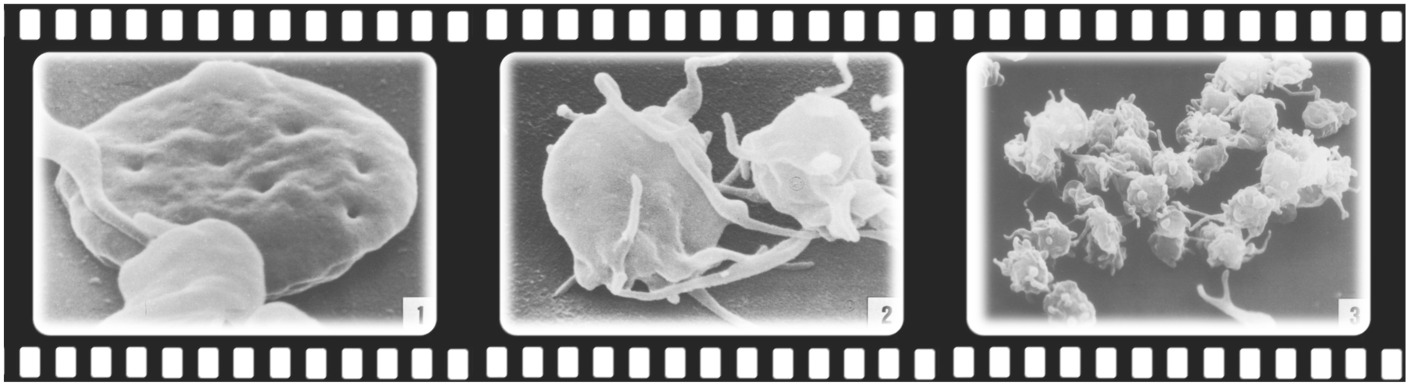
So, the objective of this regenerative procedure is to recreate the complex structure of an animal muscle using only a small number of cells. A biopsy is taken from the muscle of a live animal, and this piece of tissue is cut into small pieces to release the stem cells, which then have the ability to multiply and transform themselves into different kinds of cells depending on the medium that they are grown. As such, once these cells are cultured in a specific liquid medium, which usually contains Fetal Bovine Serum (FBS) – a serum made from the blood of a dead calf, which contains all the nutrients needed – these cells will start to grow and proliferate. Rumour goes that start-ups are already working on finding plant ingredients that can be as efficient and nutritious growing cells as FBS – which is quite expensive, and of course not animal-friendly.

Trillion of cells can be grown this way, and the marvel with nature is that these cells can naturally merge to form muscle tubes (myotubes), which can then grow to form small pieces of muscle tissue – and eventually make up a patty. To scale this process, bioreactors are used, which work similarly to the way different pharmaceutical drugs are currently produced.

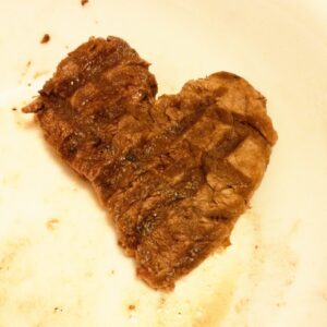
Unfortunately, the resulting patty is still far away from real muscle, which is made up of organized fibers, blood vessels, nerves, connective tissue and fat cells; as such, producing a thick piece of meat like a real steak is only a vision. “Cultivated meat” lacks the natural process of reperfusion, which is the bubbling of oxygen perfusion of blood vessels inside the meat necessary to mimic real-time nutrient diffusion. Furthermore, the richness of flavors is still significantly lower than traditional meats; with umami, bitterness and sourness still lacking due to a diminished amount of different aminoacids – the building blocks that form a protein. It’s like, such in vitro patties are only made of blue and yellow LegoTM, missing all the other colors that will make us feel fulfilled.

The problem remains that this food cannot be called vegan, because it comes originally from animal cells – whether from a chicken, or a cow, or even a frog…. And, growing meat in the lab is also not cheap at all. High amounts of liquid serum are needed to grow a couple plates of cells, not to mention grow a couple of patties to feed a family of four. Furthermore, there is a need to warm the cultured cells to mimic body temperature, so that energy needs to come from somewhere, probably coupled with CO2 emissions if fossil fuels are used. Additionally, fast growing cells are a known trigger to develop cancer – so, there needs to be a strong safety and quality assessment to make sure that cancerous cells have not developed in those in vitro meats. And another major issue is contamination: without the use of antibiotics or some other pharmaceutical means of pathogenic control, there is a high likelihood of a fully diverse & inclusive germs-party happening in those cell plates.

But truth be told, the current pressure on our food system and the increasing demands for food security, make a strong argument to find solutions for all environmental and health concerns that might arise from the field of “cultivated meat”. 

It might take some years, but it will be coming to our tables…

Would you try it?

Meatlove
Meatlove…

References:

Fountain, H. Building a $325,000 Burger. The New York Times https://www.nytimes.com/2013/05/14/science/engineering-the-325000-in-vitro-burger.html, 1 (2013). 

Chriki S, Hocquette JF. The Myth of Cultured Meat: A Review. Front Nutr. 2020 Feb 7;7:7. doi: 10.3389/fnut.2020.00007. PMID: 32118026; PMCID: PMC7020248.

Joo ST, Choi JS, Hur SJ, Kim GD, Kim CJ, Lee EY, Bakhsh A, Hwang YH. A Comparative Study on the Taste Characteristics of Satellite Cell Cultured Meat Derived from Chicken and Cattle Muscles. Food Sci Anim Resour. 2022 Jan;42(1):175-185. doi: 10.5851/kosfa.2021.e72. Epub 2022 Jan 1. PMID: 35028582; PMCID: PMC8728501.

The language of serotonin

Or, “What are they saying?

When we mention the word Serotonin (5-hydroxytryptamine, 5-HT), we immediately think of the brain and the Central Nervous System (CNS). People tend to associate serotonin to depression, or mood, or feelings of well-being1

Although that is correct, truth be told, the majority of the serotonin in the human body is actually produced in the gut. In fact, 95% of total serotonin is manufactured by the Enterochromaffin cells (or, Kulchitsky cells) in the gastro-intestinal tract (GI)2,3. These cells live next to the gut epithelium, that covers the cavity of the GI tract, playing a crucial role in the regulation of bowel movements and secretions. If you think that the gut is almost 9 meters (or 30 feet) long, then that’s a lot of cells producing serotonin. 

When in the 50’s, Betty M. Twarog and Irvine H. Page discovered that the brain produced its own serotonin4; then, the gut-made serotonin got reduced to its “Aschenputtel” origins, and relinquished to the favela quarters of the body. As such, brain-derived serotonin always got more attention than its gut-derived counterpart – like a rich vs. poor-cousin type of reputation.

Moving-on…

Platelets, also called thrombocytes, are small un-nucleated fragment of cells that, when activated, form blood clots (thrombus) and prevent bleeding. 

Electron microscopy images of circulating platelets, extracted from Zilla et al, 19875

Platelets do not make serotonin, butcan take it up as they circulate through the gut, and carry it along the blood stream6,7. As such, the serotonin produced in the intestine can be carried all over the body. As the chemical messenger serotonin is, it can influence any other cell, in whatever other location, as long as it has a serotonin receptor on it. As such, peripheral serotonin has now discovered its path back into the limelight, and recent research has strengthened the influence that gut-made serotonin has in other parts of the body, functioning as an intestinal-derived hormone. 

Once again, the “Aschenputtel” story comes into mind, but this time through its “Cinderella” version. Let’s take a look…

For example, gut-derived serotonin can directly regulate the liver and mediate liver regeneration8. In Non-Alcoholic Fatty Liver Disease (NAFLD), a group of conditions that are characterized by excessive fat accumulation in the liver and closely track the global public health problem of obesity, researchers showed that inhibiting gut-derived serotonin synthesis could resolve hepatic fat accumulation8,9.

Peripheral serotonin can also be a negative regulator of bone density, by specifically inhibiting osteoblast formation and leading to osteoporosis10 – a common feature in patients with inflammatory bowel disease (IBD). This happens through the action of a common receptor: the low-density Lipoprotein Receptor-related Protein 5(LRP5), which is expressed in both osteoblasts and enterochromaffin cells11. LRP5 inhibits the expression of an important ingredient for serotonin production (Tryptophan hydroxylase-1, Tph1); as such, when LRP5 is deficient or inactivated due to inflammation or disease, blood levels of serotonin are elevated decreasing osteoblast formation; and, consequently, reducing bone mass1,11.

Epidemiologic data suggests a role of serotonin, or Selective Serotonin-Reuptake Inhibitors (typically used as antidepressants, SSRIs) in the development of venous thrombosis12. In fact, patients with depression were reported to have higher incidences of venous thromboembolism in general13; and, the use of SSRIs is associated with an increased venous thromboembolism risk14. No wonder, serotonin and platelets are “brothers in arms”, ready to block any blood vessel along their way…. 

Serotonin and its receptors are also present in the immune system, where evidence suggests it contributes to both innate and adaptive responses. There is now clear evidence of a straight communication between the immune system, the gut and the brain via serotonin15,16.

On top of all and because we are not alone, our gut microbiota plays a critical role in regulating our colonic serotonin. Indigenous spore-forming bacteria (Sp) promote serotonin biosynthesis in our enterochromaffin cells, and with that they can significantly modulate GI movements and platelet function – together with many aspects of our physiology17,18. We now know that the microbiota colonizes the GI tract after birth, with a continuous maturation during the first years of life19. Researchers have now showed in animal models that this developing gut microbiota regulates the development of the adult enteric nervous system via intestinal serotonin networks20. What this actually means, is that the actions of our intestinal bugs during the beginning of our life are determinant for the development of our “gut brain”, our second brain. How about that?…

If we ruminate about it, when we “think” with our gut, we are actually listening to our bugs. By directly signalling our cells to produce serotonin and develop a network of neurons as soon as we are born, our gut-bugs are actually finding a way to communicate with us – the host – in the serotonin language. 

Now, we just need to understand what are they telling us… 

Beethoven’s hearing aids, Beethoven House Museum, Bonn.

References:

1          Gershon, M. D. 5-Hydroxytryptamine (serotonin) in the gastrointestinal tract. Curr Opin Endocrinol Diabetes Obes 20, 14-21, doi:10.1097/MED.0b013e32835bc703 (2013).

2          Bellono, N. W. et al. Enterochromaffin Cells Are Gut Chemosensors that Couple to Sensory Neural Pathways. Cell 170, 185-198.e116, doi:10.1016/j.cell.2017.05.034 (2017).

3          Yaghoubfar, R. et al. Modulation of serotonin signaling/metabolism by Akkermansia muciniphila and its extracellular vesicles through the gut-brain axis in mice. Scientific Reports 10, 22119, doi:10.1038/s41598-020-79171-8 (2020).

4          Twarog, B. M. & Page, I. H. Serotonin Content of Some Mammalian Tissues and Urine and a Method for Its Determination. American Journal of Physiology-Legacy Content 175, 157-161, doi:10.1152/ajplegacy.1953.175.1.157 (1953).

5          Zilla, P. et al. Scanning electron microscopy of circulating platelets reveals new aspects of platelet alteration during cardiopulmonary bypass operations. Tex Heart Inst J 14, 13-21 (1987).

6          Morrissey, J. J., Walker, M. N. & Lovenberg, W. The absence of tryptophan hydroxylase activity in blood platelets. Proc Soc Exp Biol Med 154, 496-499, doi:10.3181/00379727-154-39702 (1977).

7          Hughes, F. B. & Brodie, B. B. The mechanism of serotonin and catecholamine uptake by platelets. J Pharmacol Exp Ther 127, 96-102 (1959).

8          Wang, L. et al. Gut-Derived Serotonin Contributes to the Progression of Non-Alcoholic Steatohepatitis via the Liver HTR2A/PPARγ2 Pathway. Frontiers in Pharmacology 11, doi:10.3389/fphar.2020.00553 (2020).

9          Choi, W. et al. Serotonin signals through a gut-liver axis to regulate hepatic steatosis. Nature Communications 9, 4824, doi:10.1038/s41467-018-07287-7 (2018).

10        Lavoie, B. et al. Gut-derived serotonin contributes to bone deficits in colitis. Pharmacol Res 140, 75-84, doi:10.1016/j.phrs.2018.07.018 (2019).

11        Yadav, V. K. et al. Lrp5 controls bone formation by inhibiting serotonin synthesis in the duodenum. Cell 135, 825-837, doi:10.1016/j.cell.2008.09.059 (2008).

12        Rieder, M., Gauchel, N., Bode, C. & Duerschmied, D. Serotonin: a platelet hormone modulating cardiovascular disease. J Thromb Thrombolysis 52, 42-47, doi:10.1007/s11239-020-02331-0 (2021).

13        Takeshima, M. et al. Prevalence of Asymptomatic Venous Thromboembolism in Depressive Inpatients. Neuropsychiatr Dis Treat16, 579-587, doi:10.2147/NDT.S243308 (2020).

14        Parkin, L. et al. Antidepressants, Depression, and Venous Thromboembolism Risk: Large Prospective Study of UK Women. J Am Heart Assoc 6, doi:10.1161/jaha.116.005316 (2017).

15        Baganz, N. L. & Blakely, R. D. A dialogue between the immune system and brain, spoken in the language of serotonin. ACS Chem Neurosci 4, 48-63, doi:10.1021/cn300186b (2013).

16        Jacobson, A., Yang, D., Vella, M. & Chiu, I. M. The intestinal neuro-immune axis: crosstalk between neurons, immune cells, and microbes. Mucosal Immunology 14, 555-565, doi:10.1038/s41385-020-00368-1 (2021).

17        Yano, J. M. et al. Indigenous bacteria from the gut microbiota regulate host serotonin biosynthesis. Cell 161, 264-276, doi:10.1016/j.cell.2015.02.047 (2015).

18        Reigstad, C. S. et al. Gut microbes promote colonic serotonin production through an effect of short-chain fatty acids on enterochromaffin cells. Faseb j 29, 1395-1403, doi:10.1096/fj.14-259598 (2015).

19        Bäckhed, F. et al. Dynamics and Stabilization of the Human Gut Microbiome during the First Year of Life. Cell Host Microbe 17, 690-703, doi:10.1016/j.chom.2015.04.004 (2015).

20        De Vadder, F. et al. Gut microbiota regulates maturation of the adult enteric nervous system via enteric serotonin networks. Proc Natl Acad Sci U S A 115, 6458-6463, doi:10.1073/pnas.1720017115 (2018).

Skin microbiome: feed it right for a healthier look!

Dry skin and atopic dermatitis have been associated with changes in the variety of the skin microbiome. 

Our skin, as the largest organ in our body, has a huge array of commensal microbes that support a healthy skin barrier. One of those is Staphylococcus epidermidis, one of the most abundant bacterial species of the skin microbiome1.

This chubby mutualistic, Gram-positive, facultative anaerobe constitutes up to 90% of the aerobic resident flora of our skin, and has been associated with a healthy-looking skin2. It does not like to be lonely, and usually appears in pairs or tetrads on the surface of our skin, like a protecting biofilm.

Dry skin, for example, is associated with an increase in microbial diversity along with a decrease in microbial load in comparison to more sebaceous areas of the skin, that are usually populated by lipophilic bacteria such as Cutibacterium acnes – that tend to cause those unwanted teenager-look-a-like pimples that nobody likes…

Lactic Acid is one of the Natural Moisturizing Factors (NMF) of the skin barrier, that is essential to maintain the hydration and a slightly acidic pH of the skin surface (i.e., “acid mantle”)3. Higher lactic acid concentrations and lower skin surface pH are known to increase our epidermal renewal and promote a healthier skin. 

New in vitro data suggests that Staphylococcus epidermidis, may be one of the major sources of lactic acid in the skin1

But only if fed the right way. 

It seems that 1% colloidal oat increases Lactic Acid production by this particular bacteria species, making it rely less on simple sugars such as glucose for its metabolism; and, instead use more complex carbohydrates derived from oat.

Oatmeal-containing skin moisturisers significantly changed the metabolism of the Staphylococcus epidermidis, breaking down starch and promoting good gene expression, with an increased DNA and aminoacid synthesis, and an improved ATP metabolism.

How about that?

Bacteria on a diet makes your skin look healthier!

Next time you think about which moisturiser to buy in the drug store:  don’t forget to feed your skin microbiome it’s oatmeal!

Happy Staphys!

References:

1          Liu-Walsh, F. et al. Prebiotic Colloidal Oat Supports the Growth of Cutaneous Commensal Bacteria Including S. epidermidis and Enhances the Production of Lactic Acid. Clin Cosmet Investig Dermatol 14, 73-82, doi:10.2147/CCID.S253386 (2021).

2          Baviera, G. et al. Microbiota in healthy skin and in atopic eczema. Biomed Res Int 2014, 436921, doi:10.1155/2014/436921 (2014).

3          Thueson, D. O., Chan, E. K., Oechsli, L. M. & Hahn, G. S. The roles of pH and concentration in lactic acid-induced stimulation of epidermal turnover. Dermatol Surg 24, 641-645, doi:10.1111/j.1524-4725.1998.tb04221.x (1998).